# LAL and Gel Clot Assays for Endotoxin Detection
## Introduction to Endotoxin Detection
Endotoxins, also known as lipopolysaccharides (LPS), are toxic components found in the outer membrane of Gram-negative bacteria. Their presence in pharmaceuticals, medical devices, and other healthcare products can cause severe pyrogenic reactions in humans. Therefore, reliable endotoxin detection methods are crucial in the pharmaceutical and medical industries.
## The Limulus Amebocyte Lysate (LAL) Test
The Limulus Amebocyte Lysate (LAL) test has become the gold standard for endotoxin detection since its discovery in the 1960s. This sensitive biological assay utilizes blood cells (amebocytes) from the horseshoe crab (Limulus polyphemus) to detect and quantify endotoxins.
### How LAL Assays Work
When endotoxins come into contact with LAL reagent, they trigger a cascade of enzymatic reactions that result in clot formation. The intensity of this reaction is proportional to the amount of endotoxin present in the sample. There are three main types of LAL assays:
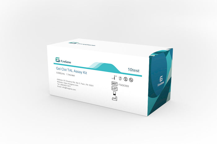
– Gel Clot Assays
– Turbidimetric Assays
– Chromogenic Assays
## Gel Clot Assays: A Traditional Approach
Among LAL methods, the gel clot assay represents the simplest and most traditional format. This qualitative or semi-quantitative method provides a visual endpoint for endotoxin detection.
### Principle of Gel Clot Assays
The gel clot technique relies on the formation of a firm gel when endotoxin activates the clotting enzyme cascade in the LAL reagent. The test involves mixing equal volumes of sample and LAL reagent, incubating the mixture, and then inverting the tube to check for clot formation.
### Advantages of Gel Clot Assays
– Simple to perform and interpret
– Requires minimal equipment
– Cost-effective compared to other LAL methods
Keyword: LAL Assays Gel Clot Assays
– Less susceptible to interference from certain sample matrices
– Provides clear pass/fail results for quality control purposes
### Limitations of Gel Clot Assays
– Less sensitive than other LAL methods (typically 0.03-0.25 EU/mL)
– Semi-quantitative at best
– Subjective interpretation of results
– Longer incubation times compared to kinetic methods
## Performing a Gel Clot Assay
The standard procedure for a gel clot assay involves several key steps:
### Sample Preparation
Proper sample preparation is crucial for accurate results. This may include dilution to overcome inhibition or enhancement effects, pH adjustment, or filtration to remove particulates.
### Test Execution
1. Prepare a series of dilutions covering the assay’s sensitivity range
2. Mix equal volumes of sample and LAL reagent in pyrogen-free tubes
3. Incubate at 37°C ± 1°C for the specified time (usually 60 minutes)
4. Gently invert each tube to check for clot formation
### Result Interpretation
A positive result is indicated by the formation of a firm gel that remains in the bottom of the tube when inverted. A negative result shows no clot formation, with the solution flowing freely when inverted.
## Applications of Gel Clot Assays
Despite the development of more advanced LAL methods, gel clot assays remain widely used for:
– Routine quality control testing of pharmaceuticals
– Raw material screening
– Medical device testing
– Water for injection (WFI) monitoring
– Validation of depyrogenation processes
## Regulatory Considerations
Gel clot assays are recognized by all major pharmacopeias (USP, EP, JP) for endotoxin testing. However, users must validate the method for their specific products and establish appropriate controls, including:
– Positive product controls
– Negative controls
– Standard endotoxin controls
– Determination of maximum valid dilution
## Future Perspectives
While gel clot assays continue to serve important roles in endotoxin detection, the field is evolving with:
– Increased automation of traditional methods